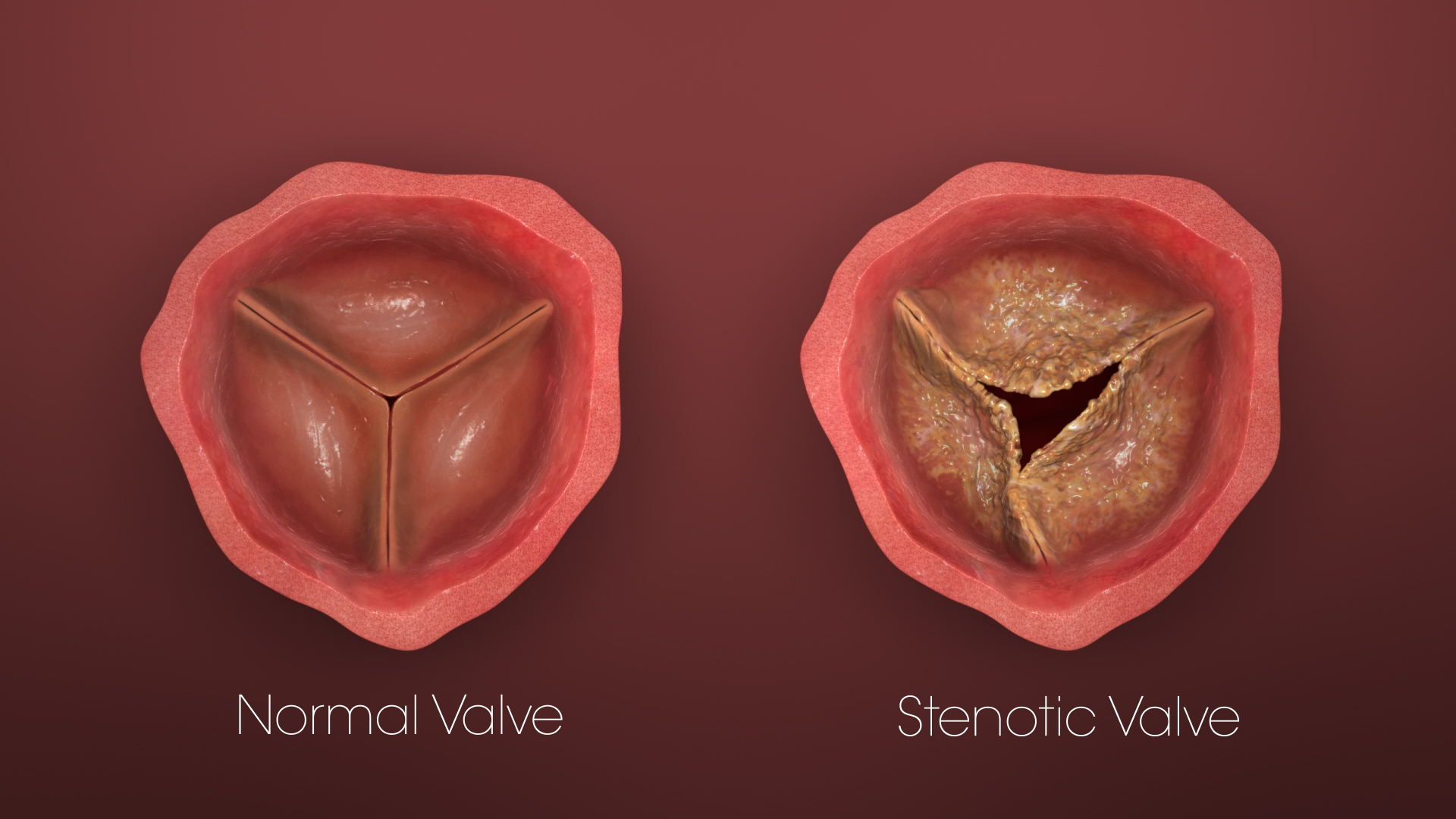
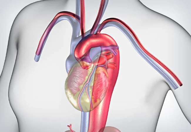
Aortic valve stenosis is a cardiovascular condition characterized by the narrowing of the aortic valve, which is responsible for regulating blood flow from the heart’s left ventricle to the aorta and the rest of the body. This narrowing restricts the blood flow, causing the heart to work harder to pump blood through the narrowed valve. It is commonly caused by the buildup of calcium deposits on the valve leaflets, leading to reduced valve opening and impaired blood circulation. Over time, aortic valve stenosis can result in symptoms such as chest pain, breathlessness, fatigue, and dizziness. Severe cases can lead to heart failure and other complications, necessitating medical intervention such as valve replacement to restore proper blood flow and alleviate symptoms.
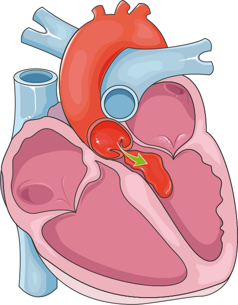
Aortic regurgitation, or aortic insufficiency, is a heart valve disorder characterized by the backflow of blood through the aortic valve into the left ventricle during the heart’s relaxation phase. This condition can arise from various causes, including valve abnormalities, aging, infections, rheumatic fever, connective tissue disorders, and aortic root dilation. Symptoms may include shortness of breath, fatigue, chest pain, palpitations, and irregular pulse. Diagnosis involves physical examinations, echocardiograms, MRIs, CT scans, and electrocardiograms.
Treatment options range from monitoring in mild cases to medical management with drugs like ACE inhibitors. In severe cases, surgical intervention may be necessary, involving either repair or replacement of the aortic valve. Procedures such as balloon valvuloplasty, a catheter-based approach, can also be employed. Lifestyle changes, including diet and exercise modifications, contribute to overall heart health.
Early detection and proper management are critical to prevent complications. Regular medical evaluations are advised for those at risk or experiencing symptoms. Aortic regurgitation requires a tailored approach, considering the severity of the condition and the individual’s overall health.
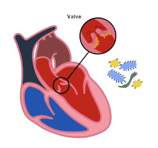
Heart valve infective endocarditis (IE) is a serious infection of the heart valves, typically caused by bacteria entering the bloodstream and settling on any one of the four heart valves. This condition can affect individuals with pre-existing heart valve issues, congenital heart defects, or those who have undergone valve replacement surgeries. The bacteria cause inflammation, leading to the formation of infected masses (vegetations) on the heart valves, compromising their function.
Symptoms include fever, fatigue, joint pain, and, in severe cases, heart failure. Diagnosis involves blood tests, echocardiograms, and sometimes other imaging studies. Timely identification is crucial as untreated IE can lead to life-threatening complications, such as heart valve damage, septic emboli, and systemic infections.
Treatment typically involves a prolonged course of antibiotics tailored to the specific infecting organism. In severe cases or when complications arise, surgical intervention may be necessary to repair or replace the infected valve.
Prevention involves maintaining good oral hygiene, promptly treating infections, and prophylactic antibiotics before certain dental or medical procedures in high-risk individuals. Early diagnosis, appropriate treatment, and preventive measures are essential in managing heart valve infective endocarditis and reducing associated risks.

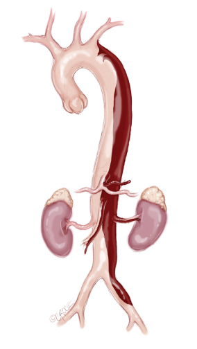
Aortic aneurysms are abnormal bulges or dilations in the aorta, the largest artery in the human body responsible for carrying oxygenated blood from the heart to the rest of the body. There are two main types of aortic aneurysms: abdominal aortic aneurysms (AAA) and thoracic aortic aneurysms (TAA).
Abdominal Aortic Aneurysms (AAA):
Most commonly occurring below the kidneys, AAA often develops silently without noticeable symptoms. Risk factors include age, male gender, tobacco use, family history, high blood pressure, and atherosclerosis. If left untreated, AAA can lead to aortic rupture, a life-threatening emergency. Diagnosis is typically made through imaging studies such as ultrasound, CT scans, or MRIs.
Thoracic Aortic Aneurysms (TAA):
Located in the chest, TAA can occur in the ascending aorta, aortic arch, or descending aorta. Causes include genetic factors, connective tissue disorders (e.g., Marfan syndrome), bicuspid aortic valve, and atherosclerosis. Symptoms might be absent or include chest or back pain, shortness of breath, or difficulty swallowing. Diagnosis involves imaging studies similar to those for AAA.
Treatment:
The management of aortic aneurysms depends on their size, location, and the individual’s overall health. Small aneurysms may be monitored regularly through imaging. Larger aneurysms or those at risk of rupture may require surgical intervention, such as open surgery or endovascular repair using stent grafts.
Prevention:
Preventive measures include lifestyle modifications like quitting smoking, managing blood pressure, and addressing underlying conditions contributing to aortic aneurysm formation. Regular check-ups, especially for individuals with risk factors, can aid in early detection and intervention.
In conclusion, aortic aneurysms pose serious health risks, and early detection is crucial for effective management. Understanding risk factors, seeking medical attention for symptoms, and adopting a heart-healthy lifestyle are key elements in preventing and managing aortic aneurysms.
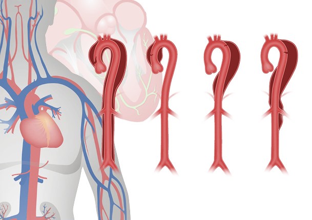
Aortic dissection is a critical medical condition characterized by the separation of the layers of the aortic wall, allowing blood to flow between them. This condition can lead to severe complications, including organ damage, stroke, or even death. Aortic dissections are classified into two main types based on the location of the tear: Stanford Type A involves the ascending aorta, and Stanford Type B involves the descending aorta.
Causes and Risk Factors:
The primary cause is often hypertension (high blood pressure), which places excessive stress on the aortic wall. Other risk factors include connective tissue disorders, genetic factors, aortic aneurysms, trauma, and certain medical conditions like Marfan syndrome.
Symptoms:
Aortic dissection typically presents with sudden, severe chest or back pain that may radiate to the neck, jaw, or arms. Other symptoms may include shortness of breath, difficulty swallowing, and neurological symptoms if the dissection affects blood flow to the brain.
Diagnosis:
Diagnosing aortic dissection is challenging due to its sudden onset and varied symptoms. Imaging studies, such as CT scans, MRIs, or echocardiograms, are crucial for confirming the diagnosis and determining the extent of the dissection.
Treatment:
Immediate medical attention is imperative in managing aortic dissection. Treatment aims to reduce blood pressure and minimize shear stress on the aorta. In Type A dissections, surgical intervention is often necessary to repair the damaged aorta. Type B dissections may be managed with medications to control blood pressure or endovascular procedures.
Prognosis and Prevention:
The prognosis depends on the extent and location of the dissection, with early diagnosis and intervention significantly improving outcomes. Prevention strategies include managing hypertension, addressing risk factors, and seeking prompt medical attention for any symptoms suggestive of aortic dissection.
In summary, aortic dissection is a life-threatening condition requiring immediate medical intervention. Early diagnosis, often through advanced imaging, and appropriate management strategies are crucial for improving outcomes and reducing the risk of complications associated with this serious cardiovascular disorder.
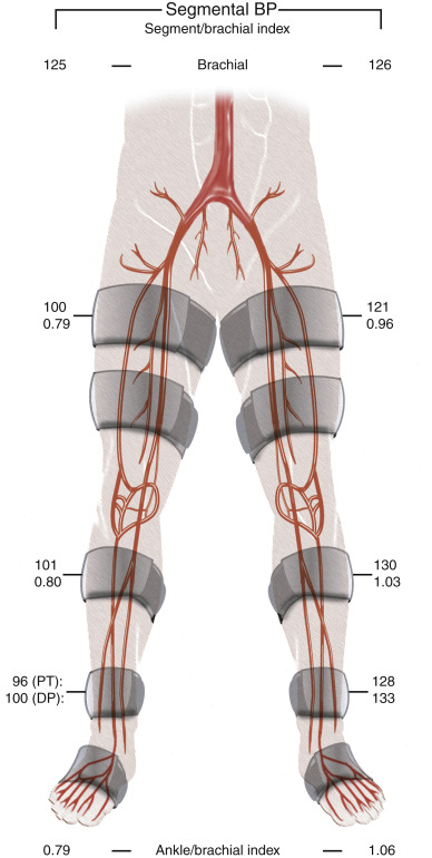
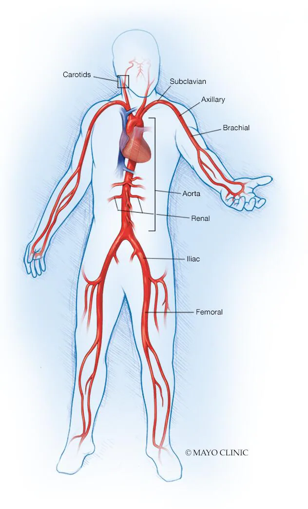
Aorta iliac occlusion refers to the blockage of blood flow in the abdominal aorta and/or iliac arteries, major vessels supplying blood to the lower extremities. This condition often results from atherosclerosis, where plaque buildup narrows or obstructs the arteries, reducing blood flow and causing various complications.
The symptoms of aorta iliac occlusion can range from intermittent claudication (pain during walking) to more severe manifestations like ischemic rest pain and even tissue necrosis. Diagnosis involves imaging studies such as angiography or Doppler ultrasound to visualize the extent and location of the blockage.
Treatment options depend on the severity and location of the occlusion. Conservative measures include lifestyle modifications, medication, and supervised exercise to manage symptoms. In more advanced cases, invasive procedures like angioplasty or stent placement may be employed to restore blood flow. Surgical interventions, such as aortoiliac bypass grafting, can be considered for complex cases.
Complications of untreated aorta iliac occlusion include the progression of symptoms, increased risk of cardiovascular events, and potential limb-threatening ischemia. The management approach aims to alleviate symptoms, improve blood flow, and prevent further complications, requiring a multidisciplinary approach involving vascular specialists, interventional radiologists, and surgeons.
Long-term care involves addressing underlying risk factors, such as hypertension, diabetes, and hyperlipidemia, to prevent disease progression and recurrence. Patient education on lifestyle modifications, regular follow-ups, and compliance with prescribed medications are crucial components of managing aorta iliac occlusion.
In conclusion, aorta iliac occlusion is a vascular condition with diverse clinical presentations, ranging from mild discomfort to severe limb-threatening ischemia. Timely diagnosis, appropriate medical management, and, when necessary, interventional or surgical interventions are essential for improving outcomes and preventing complications.
Aortic connective tissue disorders encompass a group of genetic or acquired conditions affecting the structural integrity of the aorta, the main artery that carries blood from the heart to the rest of the body. These disorders often involve abnormalities in the connective tissue components, such as collagen and elastin, leading to weakened vessel walls and an increased risk of aortic complications.
One well-known aortic connective tissue disorder is Marfan syndrome, an inherited condition characterized by mutations in the FBN1 gene, affecting fibrillin-1, a crucial protein in connective tissue. Marfan syndrome predisposes individuals to aortic dilation and dissection, where the layers of the aortic wall separate, posing life-threatening risks.
Another notable disorder is Ehlers-Danlos syndrome (EDS), a group of rare genetic disorders affecting collagen production. The vascular type of EDS specifically raises the risk of arterial and organ rupture, with a potential impact on the aorta. Additionally, Loeys-Dietz syndrome and vascular Ehlers-Danlos syndrome are other connective tissue disorders associated with aortic pathology.
Patients with aortic connective tissue disorders may present with symptoms like chest pain, shortness of breath, or back pain, which should prompt thorough cardiovascular evaluation. Diagnosis involves genetic testing, imaging studies (such as echocardiography or MRI), and clinical criteria.
Management of aortic connective tissue disorders focuses on regular monitoring to detect aortic dilation early, lifestyle modifications, and medical therapy to reduce stress on the aorta. Surgical intervention, such as aortic root replacement, may be necessary in severe cases to prevent aortic dissection or rupture.
In summary, aortic connective tissue disorders encompass a range of genetic conditions affecting the structural components of the aorta. Early diagnosis, careful monitoring, and a multidisciplinary approach involving geneticists, cardiologists, and vascular surgeons are crucial for managing these disorders and preventing life-threatening complications.
Aortoarteritis, also known as Takayasu arteritis, is a rare autoimmune vasculitis that primarily affects the large arteries, particularly the aorta and its major branches. This chronic inflammatory condition leads to vessel wall thickening, stenosis (narrowing), and sometimes aneurysm formation, affecting blood flow to various organs and tissues.
Takayasu arteritis predominantly occurs in young to middle-aged women, though it can affect individuals of any age. The exact cause is unclear, but it is believed to involve an abnormal immune response leading to inflammation and damage of the arterial walls. Genetic factors may also contribute to susceptibility.
The clinical presentation of aortoarteritis varies and can include symptoms such as fatigue, muscle pain, limb weakness, and features of systemic inflammation like fever and weight loss. As the disease progresses, patients may experience reduced pulses, blood pressure discrepancies between limbs, and, in severe cases, complications like heart failure or stroke.
Diagnosis of aortoarteritis involves a combination of clinical evaluation, blood tests to detect inflammation markers, and imaging studies such as angiography, magnetic resonance angiography (MRA), or computed tomography angiography (CTA) to visualize the arteries and assess the extent of vascular involvement.
Treatment aims to suppress inflammation, control symptoms, and prevent complications. Immunosuppressive medications, such as corticosteroids and disease-modifying anti-rheumatic drugs (DMARDs), are commonly used. In some cases, revascularization procedures or surgery may be necessary to address arterial stenosis or aneurysms.
Long-term management involves regular monitoring of disease activity, blood pressure control, and adjustments to medication based on the individual’s response. Lifestyle modifications, such as smoking cessation and maintaining a healthy diet, are essential to reduce cardiovascular risk.
In summary, aortoarteritis or Takayasu arteritis is a rare autoimmune vasculitis affecting large arteries, particularly the aorta. Early diagnosis, aggressive management of inflammation, and a multidisciplinary approach involving rheumatologists and vascular specialists are crucial for improving outcomes and preserving vascular health in individuals with this condition.
Dr. Niranjan Hiremath is a distinguished aortic expert, serving as a senior consultant and surgical lead at the Apollo Aortic Program. This program is a comprehensive unit dedicated to addressing aortic diseases, showcasing Dr. Hiremath’s expertise in the field. As a key figure in the program, he plays a pivotal role in providing specialized care for individuals with various aortic conditions.
With a focus on aortic health, Dr. Hiremath utilizes his extensive experience to lead a multidisciplinary team in the assessment, diagnosis, and management of aortic disorders. His role involves overseeing surgical interventions, collaborating with other specialists, and ensuring that patients receive personalized and effective care.
The Apollo Aortic Program, under Dr. Hiremath’s leadership, represents a commitment to delivering high-quality healthcare for individuals dealing with complex aortic issues. The program’s comprehensive approach and Dr. Hiremath’s expertise contribute to advancing patient outcomes and promoting overall cardiovascular well-being.
Dr Niranjan is also Listed on one of the top medical aggregator platform like Clinicspots